Prior to executing any radiological procedure, the points that the radiologists should take of are: Protection from the radiation, The effects of the radiations on the body, Doing Appropriately, Correct Analysis of the examination.
There is range of Radiology procedures and every examination is accomplished differently.
CT check- a diet is to be followed ordered. The individual is provided with a comparison agent by mouth, intravenously or rectally. It is a pain-free test. In this the individual is laid in a movable table as well as is glided inside a rounded device and asked to hold the breath for some time. The whole process takes a time of around 15 to thirty minutes.
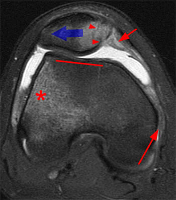
MRI scan- it is additionally a painless assessment in which is positioned under a big cylindrical magnet that has a really high magnetic field. It is a risk-free procedure Radiology treatments. Some precautions should be taken before this examination. Individuals with speed maker are not allowed to go complete this examination. The entire process takes about 1 to 2 hrs.
PET scan is increased as positron exhaust tomography. It reveals the body metabolism instead of showing the makeup. Prior to the examination the patient is administered with radioactive sugar and permitted to take remainder so that the sugar obtains distributed extensively throughout the body and afterwards moved inside the scanner to continue the procedure. It takes about 1.5 to 3 hours to complete.
Angiogram- in this examination a catheter is placed into the artery via which a comparison product is carried out. If the tests are to be taken in the early morning then the individual is not permitted to have food as well as beverage water after twelve o'clock at night. This test is a painless one and takes close to regarding 2 hrs.
Ultrasonography- in this acoustic wave with high frequency are use to picture inside the body and after that received by the transducer which is considered as an photo in the monitor. USG is of numerous types. Pelvic stomach, renal etc. In pelvic USG the patient is asked to consume 30-to 45 oz of water before the test. This test is likewise pain-free.
Radiology technologists take xrays and carry out nonradioactive materials into clients' blood streams for analysis purposes. Some concentrate on analysis imaging modern technologies, such as computerized tomography (CT) and also magnetic resonance imaging (MRI). Radiologic engineers and also specialists, also described as radiographers, generate xray films (radiographs) of parts of the human body for use in diagnosing clinical troubles.
They prepare patients for radiology tests by discussing the treatment, removing write-ups through which xrays can not pass as well as positioning people so that the parts of the body can be properly radiographed.To prevent unneeded radiation direct exposure, these workers border the subjected location with radiation protection devices, such as lead shields, or limit the dimension of the xray beam with collimation.
Radiology technologists setting radiographic devices at the right angle and also elevation over the proper area of a client's body. Utilizing tools similar to a determining tape, they might determine the density of the area to be x-rayed and also established controls on the xray machine to produce radiographs of the ideal thickness, detail, as well as comparison. They position the x ray movie under the part of the client's body to be checked out as well as make the direct exposure. They after that get rid of the film as well as create it.
After inspecting the film for high quality, the rad tech will certainly send it to the radiologist for interpretation. The patient is launched as well as told to anticipate the results.
Radiology is an additional type of clinical specialty which is used ti obtain photos of various parts of the body to spot as well as deal with conditions. Different imaging strategies are utilized by the radiologists as well as one of the most crucial among them are X-RAY, USG, CT Scan, nuclear medicine, PET and also MRI.
There are different sorts of Radiology techniques which are stated as under:-.
X-Rays- it is likewise referred to as radiographs. There are generated by passing x-rays via the patient's body which then obtains guided to a recording device and also additional established as an image. One of the most generally pre-owned form of imaging is the Silver Containing movies which is currently changed by Digital radiography. As a result of its accessibility as well as affordable rates is one of the most prescribed test given by the medical professionals.
Fluoroscopy- Angiography or Fluoroscopy are the special form of an x-ray applications. In this a screen and an intensifier is made use of which assist in the formation of the picture both this things are attached to a close circuit television. The patient as provided with contrasting agents to set apart in between the tissues. It is typically used to identify growths or cysts.
Interventional radiology- it is primarily made use of to identify and treat peripheral vascular conditions, Inferior vena cave filter positioning, gastrostomy tube positionings, biliary stents and hepatic interventions in a minimally invasive technique.
Computed Tomography- X-rays is made use of additionally with formulas to take image of the body. It is made use of for detecting urgent circumstances such as hemorrhage, clots in the arteries of the lungs, appendicitis, as well as curing kidney stones.
Ultrasound-it is utilized to picture the unborn child, kidney rock, spleentomegaly and so on it used the high frequency sound waves to discover the problems.
Magnetic Resonance Imaging- it utilizes electromagnetic fields to locate the core of the atom within the tissues, after that utilizes a radio signals to produce disturbance in the axis of rotation of core and also observes the superhigh frequency signal generated As well as none the less are the nuclear medicines imaging which are carried out into the people consisting of products which have the affinity for cells classified with radioactive tracer.
Ideally, radiology services need to be available 24/7 in clinical facilities for the fast analysis of exams and prompt treatment of medical problems. This is especially vital for emergency scenarios where time is of the essence.
Regrettably, this isn't constantly the instance - specifically for smaller health centers, centers or methods. The arrival of teleradiology has actually made this feasible for these organizations as well as people, permitting them to supply faster, top quality individual care.
Healthcare facility emergency rooms, surgical wings, as well as other highly vital clinical treatment atmospheres regularly require radiological pictures taken as soon as possible for individuals who deal with mishaps or serious conditions that emerge all of a sudden. With teleradiology, medical professionals have the ability to enlist expert radiology services right away to assist secure a medical diagnosis.
Certain companies are devoted exclusively to offering such services as well as use permanent radiologists that are available round-the-clock. Such services can consist of a variety of specialties and also subspecialties, ranging from body imaging, to pediatric radiology, to cardiovascular imaging. Radiologists use the most recent picture archiving and also communications system to get, examine as well as analyze radiological pictures, such as X-rays, MRIs and CT scans.
With the best modern technology as well as high-speed Net, radiologists can do these and generate both preliminary and also last records from their houses, despite the time or day. In some business, reports can even be made available in as soon as 30 minutes. Teleradiology has actually paved the way for more rapid medical diagnosis and also treatment.
Speed, dependability and also adaptability have actually made this practice gain boosting popularity among clinical facilities across the nation. Because when it concerns individual treatment, there's no time to waste.
Numerous techniques are utilized to picture organs either directly or indirectly, Vistas of clinical imaging have broadened explosively in recent years and also are still developing rapidly.
One of the most classic and well developed approach of imaging is radiology, which uses X-rays. Shadows cast on the photosensitive film by various cells vary in thickness as well as this principle is sued in interpreting the radiographs. Different methods like ordinary radiography, contrast radiography as well as tomography are utilized. Radiological imaging offers details about physiological as well as architectural changes in an body organ, e.g, foreign bodies in the bronchi, combination of the lungs, cardiac enhancement, abnormalities of bones, etc. Both the anatomic irregularities and also physiological features can be examined by methods utilizing contrast radiography, e.g, barium ingest, barium dish follow through, cholecystography, comparison urography, etc. Angiography elegantly exposes the vascular supply of an body organ. Aside from imagining occlusion as well as aneurysms, the vascular pattern provides indirect evidence of lumps, area occupying lesions and likewise the practical state of the body organ. Angiography has actually been extensively applied in Cardiovascular, neurological, kidney, hepatic, and also other problems. Angiography has actually been made use of with various other methods like computerized tomography to boost the resolution of information further. Angiography has been put on the arteries, blood vessels as well as lymphatics.
A new advancement in the field is interventional radiology in which investigatory or healing treatments are done under radiological control. The technique is highly innovative, demanding extremely fantastic ability and also perfect team job. A few classic instances of interventional radiography are endoscopic backward cholangiopancreatography (ERCP) with removal of pancreatic or biliary calculi; kidney artery dilation via a kidney artery catheter and also alleviation of coronary occlusion utilizing a balloon catheter in the coronary artery.
Radiology is still the common strategy of imaging considering that this examination answers the majority of the questions. Moreover its universal schedule and reasonably affordable have aided to make it the most acceptable investigation. Though traditional readiography is noninvasive, contrast researches are intrusive in varying levels. The direct exposure to diagnostic X-rays, though small quantitatively, contributes to cumulative irradiation obtained by the subject. It is well-known that irradiation of the fetus in utero, particularly during early pregnancy can be hazardous to the infant. So also duplicated radiographic studies can provide cumulative toxicity as a result of X-rays. Though the dosage and the area of direct exposure have been considerably reduced in https://codyeamt821.skyrock.com/3337177286-Responsible-For-A-Diagnostic-Centre-Budget-10-Terrible-Ways-To-Spend.html modern-day equipments, this threat must not be ignored and radiological researches must be embarked on just if appropriately suggested. Several radiological methods are supplemented by the newer imaging techniques like Ultrasonography, isotope scanning, computerized tomography as well as nuclear magnetic resonance.